Home
/ Upper Thigh Cross Sectional Anatomy : Anatomy Atlases Atlas Of Human Anatomy In Cross Section Section 7 Lower Limb : Harry benjamin laing, mrcs, ortho m8, frcs(tr and orth) tutorials
Upper Thigh Cross Sectional Anatomy : Anatomy Atlases Atlas Of Human Anatomy In Cross Section Section 7 Lower Limb : Harry benjamin laing, mrcs, ortho m8, frcs(tr and orth) tutorials
Upper Thigh Cross Sectional Anatomy : Anatomy Atlases Atlas Of Human Anatomy In Cross Section Section 7 Lower Limb : Harry benjamin laing, mrcs, ortho m8, frcs(tr and orth) tutorials. Anatomy of the thigh and leg the thigh is best described in terms of compartmental anatomy, and is composed of anterior, posterior, and medial (adductor) compartments. It consists of three muscle compartments (anterior, posterior, medial) which create movement by acting on the femur bone. On the full length video, we will explore them on cross section. 9 public playlist include this case. Tendons are cords made of tough tissue, and they work as special connector pieces between bone and muscle.
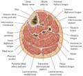
Muscles of the lower limb; Cross sectional anatomy browser includes There are many structures, like muscles and bones that can be found in the upper arm region. A cross section of the thigh vectorized and adapted from figure 432 from the 1918 grays anatomy. Each compartment has a distinct innervation and function.
Anatomy Atlases Atlas Of Human Anatomy In Cross Section Section 7 Lower Limb from www.anatomyatlases.org Cross sectional anatomy of the hip : Cross sectional anatomy of distal lower leg. Online mri & ct sectional anatomy kenneth k. Knee and lower leg clinical gate. Each compartment has a distinct innervation and function. Harry benjamin laing, mrcs, ortho m8, frcs(tr and orth) tutorials Use the mouse scroll wheel to move the images up and down alternatively use the tiny arrows (>>) on both side of the image to move the images.>>) on both side of the image to move the images. Thigh muscle anatomy mri thigh muscles cross sectional anatomy radiology case picture of thigh muscle anatomy mri thigh muscles cross sectional anatomy radiology case.
Harry benjamin laing, mrcs, ortho m8, frcs(tr and orth) tutorials
Welcome to online mri & ct sectional anatomy. Each compartment has a distinct innervation and function. A review reg anesth pain med. It consists of three muscle compartments (anterior, posterior, medial) which create movement by acting on the femur bone. Diagnosis not applicable diagnosis not applicable. Meanwhile, the vastus lateralis is on the side of the thigh, while the vastus intermedius is hidden below the rectus femoris(5). Please email baodo at stanford.edu Not only the fascia seems to be more dilative also the. Instant anatomy is a specialised web site for you to learn all about human anatomy of the body with diagrams, podcasts and revision questions Together, the lumbar and sacral plexus supply innervation to the lower extremity. Knee and lower leg clinical gate. The cross sectional human anatomic atlas of the lower limb is an interactive tool based on mr axial images of the human leg. Like the biceps brachii in the arm, the biceps femoris muscle has two heads.
Our first stop is the thigh. Cross sectional anatomy of the hip : The thigh is the thickest portion of the lower extremity, located between the hip and knee. A review reg anesth pain med. Knee and lower leg clinical gate.
Cross Sectional Anatomy Of The Thigh Demonstrating The Anterior Download Scientific Diagram from www.researchgate.net Stanford bone tumor ddx | iss/ssr msk lectures | search ocad cases | stanford virtual readouts stanford msk mri atlas has served over 1,000,000 pages to users in over 100 countries. The muscles of the lower limb are numerous and complex. The four muscles all extend the lower leg. On the full length video, we will explore them on cross section. Instant anatomy is a specialised web site for you to learn all about human anatomy of the body with diagrams podcasts and revision questions. Cross sectional anatomy of distal lower leg. Together, the lumbar and sacral plexus supply innervation to the lower extremity. Not only the fascia seems to be more dilative also the.
Serial cross sections variant image id:
A cross section of the thigh vectorized and adapted from figure 432 from the 1918 grays anatomy. The greater saphenous vein courses along the _____ side of the thigh/calf. Our first stop is the thigh. This mri wrist coronal cross sectional anatomy tool is absolutely free to use. Knee and lower leg clinical gate. How you will use this image and then you will be able to add this image to your shopping basket. Welcome to online mri & ct sectional anatomy. The rectus femoris is located in the center of the thigh, while the vastus medialis is in the middle of the said body part. Cross sectional anatomy of the hip : Cross sectional anatomy browser includes 9 public playlist include this case. Support radiopaedia and see fewer ads. All motor innervation to the posterior thigh derives from the tibial division of the sciatic nerve except for the short head of the biceps femoris.
The musculature of the thigh can be split into three sections; The thigh is the thickest portion of the lower extremity, located between the hip and knee. There are many structures, like muscles and bones that can be found in the upper arm region. Diagnosis not applicable diagnosis not applicable. Cross sectional anatomy browser includes
Atlas Of Anatomy Vol 1 General Anatomy Cross Sectional Anatomy Of The Thigh Leg And Foot from thiemelegacy.blob.core.windows.net The book is designed to help novices acquire pattern recognition skills to resolve. Instant anatomy is a specialised web site for you to learn all about human anatomy of the body with diagrams podcasts and revision questions. A review reg anesth pain med. Stanford bone tumor ddx | iss/ssr msk lectures | search ocad cases | stanford virtual readouts stanford msk mri atlas has served over 1,000,000 pages to users in over 100 countries. There are many structures, like muscles and bones that can be found in the upper arm region. The greater saphenous vein courses along the _____ side of the thigh/calf. This mri wrist coronal cross sectional anatomy tool is absolutely free to use. Use the mouse scroll wheel to move the images up and down alternatively use the tiny arrows ( >> ) on both side of the image to move the images.
The cross sectional human anatomic atlas of the lower limb is an interactive tool based on mr axial images of the human leg.
The rectus femoris is located in the center of the thigh, while the vastus medialis is in the middle of the said body part. Diagnosis not applicable diagnosis not applicable. Arteries lower leg this mri shoulder axial cross sectional anatomy tool is absolutely free to use. Cross sectional anatomy browser includes Use the mouse scroll wheel to move the images up and down alternatively use the tiny arrows ( >> ) on both side of the image to move the images. The musculature of the thigh can be split into three sections; Anatomy of the thigh and leg the thigh is best described in terms of compartmental anatomy, and is composed of anterior, posterior, and medial (adductor) compartments. The book is designed to help novices acquire pattern recognition skills to resolve. All motor innervation to the posterior thigh derives from the tibial division of the sciatic nerve except for the short head of the biceps femoris. A cross section of the thigh vectorized and adapted from figure 432 from the 1918 grays anatomy. Support radiopaedia and see fewer ads. Muscles of the lower limb; The greater saphenous vein courses along the _____ side of the thigh/calf.

 tutorials)